Almost everyone has a skin lesion of some type. Although most can easily be ignored, but some may be more conspicuous and can detract from your skin’s natural beauty.
Types of benign skin lesions:
- Pigmented lesions
- Skin colored lesions
Benign Pigmented Lesions
Benign pigmented skin lesions are an extremely common dermatological problem. They are formed by melanin concentration spots in the skin and can be of any size or shape and may also be slightly elevated on the skin’s surface.
Patients consider a number of these lesions cosmetically undesirable because of their location, color, size, or other clinical features.
Most common benign pigmented lesions:
- Freckles
- Solar lentignes or Liver spots
- Cafe au lait spots
- Seborrhoeic keratosis
Laser treatment for benign pigmented lesions
Prior to the introduction of lasers, epidermal pigmented lesions have been treated using a destructive method to remove the epidermis containing the lesion. Common destructive methods have included cryotherapy, dermabrasion, electrodessication, and chemical peeling.
Because such methods are essentially nonspecific in their destruction of the epidermis, side effects such as permanent hypopigmentation, atrophy, scarring, and changes in the skin surface texture may result.
Therefore, treatment of pigmented lesions by selectively targeting the pigment-containing cells is preferable.
Advances in laser technology have provided a safe and effective alternative for the treatment of many types of benign pigmented skin lesions. Selective destruction of pigmented lesions has been best achieved via selective photothermolysis using pigment-specific lasers.
Through the selection of laser parameters (i.e. wavelength, pulse duration, fluency, and spot size), the pigment bearing cell can be targeted directly enabling selective destruction of the offending pigment while preserving surrounding normal skin and minimizing cosmetically unacceptable side effects.
The procedure
Patients should remove all make-up and sun block from the area to be treated. The area around the nevus should also be shaved prior to treatment. A layer of anesthetic gel is applied on the treatment area. When the skin is numbed the laser moves gently over the area. The laser breaks the melanin into micro-particles, lightening and removing the dark spot.
After the procedure there may be some stinging, but it can be alleviated by the application of ice packs.
The treated area should be gently washed at least twice daily with soap and water. In the event the epidermis has been removed, an ointment, such as Aquaphor or aloe vera gel, and an occlusive dressing can be applied. Patients should avoid excessive sun exposure and use a zinc-oxide-based sunscreen with an SPF 20 or higher after treatment to avoid the chance of hyperpigmentation.
Freckles
Freckles are flat circular spots on the skin, about the size of a sesame seed. They usually develop in bilateral symmetry around the nose and lower eyes. Freckles may be tan, light-brown or black in color.
Freckles are most commonly the result of genetics or sun exposure.
They may appear at age 10, and are found more often in girls in their teens and twenties. Freckles may develop darker after repeated exposure to sunlight in the summer.
Treatment
Freckles can be removed by Nd YAG laser or CO2 laser treatments.
The Nd YAG laser targets only the cells, that contain the excess pigment, leaving normal, healthy skin unharmed. Melanin- the pigment that causes freckles to be darker than surrounding skin, is broken up causing the freckles to become lighter and disappear.
Fractional CO2 laser also improves freckles. Fractional laser creates tiny micro holes in the skin, that heal over rejuvenating and tightening the skin and minimizing the freckles. More about the procedure here.
Solar lentigines (liver spots or sun spots)
Solar lentigines or sun spots are small pigmented lesions, that usually appear on areas of the body that are frequently exposed to the sun such as the face, hands, shoulders and arms. They can be of any color from light brown to black and are associated with aging and exposure to ultraviolet radiation from the sun.
They are sometimes called liver spots because they were once incorrectly believed to be caused by liver problems, but they are unrelated to the liver.
Solar lentigines differ from freckles as freckles have a relatively normal number of pigment bearing cells but an increased amount of melanin pigment. A lentigo has an increased number of melanocytes. Freckles will increase in number and darkness with sunlight exposure, whereas lentigines will stay stable in their color regardless of sunlight exposure.
Treatment
Liver spots develop in three types; epidermal, dermis and complex, and an appropriate treatment should be applied to the type. It is important to remember, that early treatment can prevent further appearance and darkening of liver spots.
Sun spots respond well to laser treatments and can be removed with Nd YAG laser to target the excess pigment bearing cells.
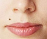
Café au lait spots or Café au lait macules
Cafe au lait spots are light brown, flat and larger in size than other types of pigmented lesions that are usually located on the trunk. The size of the lesions varies Cafe au lait spots are usually classified as a type of birthmark because they exist at birth. However, some may appear with age or with the development of the disease neurofibromatosis. Cafe au lait spots that appear as an indication of neurofibromatosis are generally larger and appear at a greater frequency on the body
Treatment
Cafe au lait spots can be treated with the Nd YAG laser, that targets only the cells with excess pigment, leaving normal, healthy skin unharmed or with Fractional CO2 laser.
Seborrhoeic keratosis
Seborrhoeic keratosis are wart like lesions of various colors that “look stuck on” like a scab from a healing wound.. Seborrhoeic keratosis are most likely caused by hereditary factors and changes in the skin growth factor.
Treatment
The treatment of Seborrhoeic keratosis depends on their pigmentation. Darker lesions can be treated with the Nd YAG laser, that targets only the cells with excess pigment, leaving normal, healthy skin unharmed. Lighter lesions respond well to treatments with Fractional CO2 laser.
Skin colored lesions
Most common benign Skin Lesions:
- Warts: Benign skin growths that are caused by human papillomavirus (HPV). They appear as small rough papules (2-10 mm) that can resemble a cauliflower or a solid blister, typically appearing on hands and feet but can also grow on other locations. Warts are contagious on contact.
- Papillomas: Common benign skin tumors that are also caused by a type of human papillomavirus (HPV). Papillomas resemble soft, skin colored, finger like growths (0,2-10mm in diameter) usually situated on face, neck, armpits and groin. They have the tendency to spread and affect large areas if untreated. Papillomas are contagious on contact.
- Condylomas: Condylomas are pointed, warty formations in the form of soft lobule growths that are also caused by the (HPV). They are located in the anogenital region: on the mucous membrane of urethra or rectum, in the perianal region. Sometimes Condylomas can grow fast to remarkable sizes. Condylomas are sexually transmitted.
- Syringomas are harmless sweat duct tumors, typically found clustered on eyelids, although they may also be found in the armpits, umbilicus, or vulva. They are skin-colored or yellowish firm rounded bumps, sized 1-3 mm in diameter.
Treatment
Laser “vaporization“ of skin lesions can quickly and safely remove: Warts, Syringiomas, Colloid Milium, Xanthelasma, and other benign skin lesions.
The method is painless, and leaves only a minimal mark on the patient’s skin surface.
The benefits of laser treatment:
- Controlled depth of influence
- Less damage to surrounding tissue
- The lesion is removed precisely in its borders
- No wounds that need closing
- Easy care after the procedure
- Minimal risk of complications